BK Polyomavirus (BKV)
BK Polyomavirus (BKV) is an icosahedral DNA virus with a size of 45-50 nm. BKV was first described in 1971 following the finding of inclusion‐bearing in the urine of a kidney transplant recipient. This virus, along with John Cunningham virus (JC) and Simian virus 40 (SV40), is a member of the polyomavirus (PV) family. BKV is an important risk factor for BKV-associated diseases such as BKV Nephropathy (BKVN) and the failure of Kidney transplantation.
BKV infects most of the world’s population, often at a young age. This type of infection is inactive and without clinical symptoms. BKV is generally transmitted through respiratory secretions. After the first infection, a type of inactive infection is established in kidney cells and Uroepithelium, and BKV induces inclusion bodies with the size of 40-45 nm in renal tubular cells. Although BKV infection is inactive and without clinical symptoms in most people; In immunocompromised or immunosuppressed patients people, BKV is reactivated and replicates. BKV reactivation causes a series of effects including The lysis of tubular cells and the secretion of BKV in the urine. Activated BKV will lead to problems such as nephropathy and graft failure. One of the main reasons for graft failure is the immune rejection of the transplanted organ, which has various causes, including BKV-related diseases.
BKV-related Diseases are usually observed in kidney and hematopoietic stem cell receiving patients. BKV nephropathy or BKVN is the most important type of BKV-related Disease. BKVN is defined as a viral plasma load of more than 10,000 Copies/ml continuously over 28 days. In terms of histology, BKVN is classified into three levels: A, B, and C. At stage A, the strong cytotoxic effect is followed by a sustained tubulointerstitial inflammation (stage B); this condition leads to tubular atrophy and interstitial fibrosis (stage C). BKVN occurs in 10% of organ transplanted recipients, especially in cases where the recipient’s and donor’s blood groups are incompatible, with the possibility of transplant rejection between 10-80%. The main reason for the reactivation of BKV in these conditions is the immunosuppressive treatments after the transplantation.
The BKV seroprevalence is estimated to be more than 80% in immunocompetent people. The seroprevalence seems to decrease with age. Intermittent viral shedding has been reported in 7-20% of urine samples in healthy individuals, and this rate is higher in immunocompromised individuals. Four genotypes of BKV have been identified in kidney transplanted patients; genotype I is prevalent worldwide, genotype III is more frequent in Africa, and genotype IV is prevalent in Asia and parts of Europe.
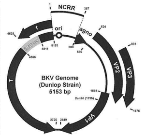
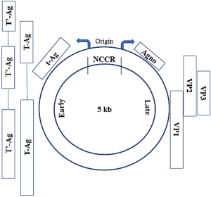
BKV genome is circular double-stranded DNA with a size of 5 kb including three regions; Early (which encode st and LT antigens), Late (which encode Vp1, Vp2, and Vp3 capsid proteins, agnoprotein, and microRNAs), and non-coding control region (NCCR). Based on Vp1 and NCCR polymorphisms; BKV strains are classified into 6 genotypes (I-VI).

| 
|
The NCCR control region contains the origin of replication (ori) along with the promoters of the Early and Late genes. The difference in the NCCR region makes a distinction between two different types of BKV, which include archetype and rearranged BKV. The archetype is the transmissible state of the virus that causes an inactive and asymptomatic infection in the body. Rearranged BKV has deletion and duplication in NCCR and is the type associated with the BKV- related disease. Transcription from the Early region leads to the production of st and LT antigens. LT has a regulatory role in the expression of Late genes and due to its helicase activity, it also plays a role in virus DNA replication. Late genes expression lead to the production of structural proteins (Vp1, Vp2, Vp3, and agnoprotein). Vp1 is the most important structural protein; the BKV capsid structure is obtained from the arrangement of 360 Vp1 molecules together in a pentameric structure. Vp2 and Vp3 proteins are attached to the inner surface of the capsid. Agnoprotein plays an important role in the viral infectivity cycle and helps release and spread the replicated viruses from infected cells.
BKV enters the cell through the binding of VP1 to its receptors on the cell surface. After endocytosis and transfer of the virus to the endoplasmic reticulum, the virus is uncoated and its genome enters the nucleus. The virus genome is replicated and translated into the cell nucleus. By expressing Late genes, structural proteins form the capsid, which may be released from the infected cell in an active manner.
Among the diagnostic methods, renal biopsy is the gold standard for diagnosing BKV-related nephropathy; However, this method is time-consuming, invasive, and dependent on the user’s skill. Also, due to the focal nature of the infection and the tendency of the primary infection to reach the deeper parts of the kidney, it is usually not possible to diagnose nephropathy using this method. Currently, cytological methods of BKV detection in urine samples are not performed due to the low specificity of this method. The ease and availability of virus DNA detection methods using the quantitative Real-Time PCR or qPCR technique have made this technique the main method of BKV diagnosis. The two organizations, Kidney Diseases Improving Global Outcomes (KDIGO) and the American Society of Transplantation (AST) guidelines also recommend BKV screening using the qPCR technique in all renal transplants.
Quantitative Real-Time PCR or qPCR is a technique that uses PCR to amplify desired gene targets. This method uses fluorescent molecules to detect amplified products. During the reaction and amplification of nucleic acid, the amount of fluorescence radiation increases; As a result, it is possible to real-time analysis of PCR products quantitatively. Using non-specific fluorescent dyes (such as Cyber green) or using specific probes labeled with fluorescent dyes (such as TaqMan probes) are two common methods of viral genome detection. As a diagnostic tool, probe-based Real-Time PCR is used in the rapid and accurate detection of infectious viruses’ nucleic acid.
BKV infection can be detected using qPCR in plasma or urine samples; The test result is considered positive when the number of viral copies is more than 107 copies/ml in urine and 104 copies/ml in plasma. Similar results should also be repeated within 4 weeks.
BKV DNA Detection using qPCR cannot distinguish active viruses from inactive ones; For this reason, the detection of viral mRNA, especially VP1 mRNA, has been considered as an indicator of active viruses in the nephropathy diagnosis. Also, the detection of viral microRNAs or miRNAs using the qRT-PCR technique has been considered a non-invasive method in BKV diagnosis. Bkv-miR-B13p and bkv-miR-B1-5p are two miRNAs expressed by the virus and involved in the regulation of replication and immune evasion. These two miRNAs have been used in active BKV infection detection using qRT-PCR.
There is no specific treatment for BKV-related Diseases and transplant rejection, and usually, improvement is based on reducing immunosuppressive drugs. Increased care and early detection of virus infection can bring favorable results.

